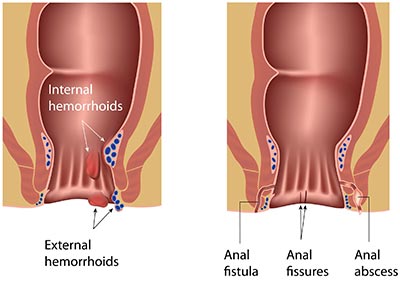
Acute rectal bleeding, also known as lower gastrointestinal (GI) bleeding, is the loss of fresh blood from the colon.
Blood in stool can result from a number of causes usually categorized as:
- Anatomic or diverticulosis
- Anflammatory such as infections and inflammatory bowel disease
- Vascular such as angiodysplasia, radiation induced causes, and ischemic,
- And neoplastic causes.

Rectal Bleeding Management
Acute lower GI bleeding can also result from polypectomy and other similar therapeutic interventions. This article looks at the clinical manifestations of acute rectal bleeding, its diagnosis, management, and treatment.
Clinical Manifestation
Patients with lower GI bleeding usually report hematochezia, which refers to the passage of bright red or maroon blood or clots from the rectum. Blood from the left side of the colon is usually bright red in color while the blood coming from the right side has a darker or maroon appearance and it may be mixed with the patient’s stool. It’s very rare for patients bleeding from the right side of the colon to have melena.
Patients with acute bleeding from the anus should also have normocytic red blood cells. Chronic lower GI bleeding can also be a sign of iron or microcytic red blood cells deficiency. Contrary to upper GI bleeding, a patient with acute rectal bleeding while showing a normal renal perfusion should also show signs of normal blood urea nitrogen.
Initial Evaluation and Management of Acute Lower GI Bleeding

Rectal Bleeding Treatment
Initial evaluation of a patient with signs of acute rectal bleeding should run parallel with the management of the condition. The goal of the initial evaluation is to find out if the blood is originating from the lower GI tract, check for the severity of the bleeding, find the most appropriate setting for the patient, and implement the necessary supportive measures before initiating resuscitation. After completing these initial steps, other diagnostic studies such as colonoscopy can follow based on the American College of Gastroenterology 2016 guidelines.
Initial Evaluation
There are several steps to follow in the initial evaluation which include getting the patient’s history, doing a physical examination, conducting laboratory tests, and if necessary doing a nasogastric lavage or upper endoscopy. The goal of this evaluation is to confirm that the bleeding is not coming from the upper gastrointestinal tract and that the condition can be effectively managed.
- History
It’s important to get the patient’s medical history at the initial stage. Find out if the patient has had previous episodes of lower or upper GI bleeding. This will help you identify potential causes for the bleeding that may influence further management of the condition. Inquire about the patient’s medication use focusing on drugs or agents that could potentially cause bleeding or impair coagulation such as anticoagulants, non-steroidal anti-inflammatory agents, and any other antiplatelet agent the patient may have used in the past.
- Physical Examination
Physical examination involves the assessment of the patient’s hemodynamic stability and his or her stool to check for the presence of melena or hematochezia. Check if the patient has abdominal pain which could be a sign of an inflammatory bleeding source, for example infectious colitis, ischemic, or a perforation such as a perforated peptic ulcer.
- Laboratory Tests
Laboratory tests done on the patient should include serum chemistries, liver tests, complete blood count, and coagulation tests. The patient’s hemoglobin level should be checked after every 2 to 8 hours depending on how severe the bleeding is.
Management
The first step in the management of a patient suspected to have acute rectum bleeding is to find the most appropriate setting for the patient, which could be outpatient, inpatient, or intensive care unit. A colonoscopy and endoscopy doctor in South Florida will also provide the patient with general supportive measures at this stage which can include adequate intravenous access and oxygen supply. Management will also involve giving the patient the necessary blood and fluid product resuscitation and management of anti-coagulants, coagulopathies, and anti-platelet agents.
A patient with high-risk features such as hemodynamic instability usually characterized by orthostatic hypotension and shock, comorbid illness, or persistent bleeding should be transferred to intensive care unit for closer observation and the necessary therapeutic intervention. Close observation for a high-risk patient in the ICU will include electrocardiogram monitoring, pulse oximetry, and automated monitoring of the patient’s blood pressure.
Some of the key features used to categorize high risk and low risk patients include hemodynamic stability, advanced age, comorbid illness, persistent bleeding, rectal bleeding in a patient admitted for another illness, a previous history of angiodysplasia or diverticulosis bleeding, aspirin use, a non-tender abdomen, a high-level of blood urea nitrogen, anemia, and an abnormal count of white blood cells.
Management should also include the following:
- General supportive measures such as supplemental oxygen through the nasal cannula rather than the mouth just in case an upper endoscopy is urgently needed.
- Fluid resuscitation and stabilization with intravenous fluids.
- Blood transfusion. Young patients who don’t have comorbid illness may not require blood transfusion unless their hemoglobin levels drop below 7 g/dL. Older patients will require blood transfusion to maintain higher hemoglobin levels of 9g/dL and above.
Diagnosis

Rectal Bleeding treatment in Florida
After you have ruled out upper GI as the source of the bleeding, the first step of diagnosis is colonoscopy. Other diagnostic studies usually conducted on patients with lower GI bleeding include radionuclide imaging, mesenteric angiography, and computed tomographic or CT angiography.
Colonoscopy in high-risk patients or those with ongoing bleeding should be done within 24 hours after adequate bowel preparation.
Treatment of Lower GI Bleeding
The treatment of the bleeding site focuses on the source of the bleeding. The bleeding can be controlled in most cases by specific therapies applied during colonoscopy or angiography. Surgery is rarely required in most cases but if it has to be done, it will be important to first determine the source of the bleeding and what has caused it.
Conclusion
The treatment of rectal hemorrhoids and bleeding starts with an initial evaluation which includes the patient’s medical history, an overall physical examination, and laboratory tests. In some cases, it may be necessary to conduct upper endoscopy or nasogastric lavage to confirm the source of bleeding is actually in the lower GI tract. The actual treatment will depend on the source and cause of bleeding. In most cases treatment is done during the colonoscopy or angiography phase. Most rectal bleeding patients with red stool or black stool symptoms rarely require surgery.
The post Rectal Bleeding Diagnosis, Management, and Treatment appeared first on Gastroenterologists In Florida.
from
https://gastroinflorida.com/blog/rectal-bleeding-diagnosis-management-and-treatment/

No comments:
Post a Comment